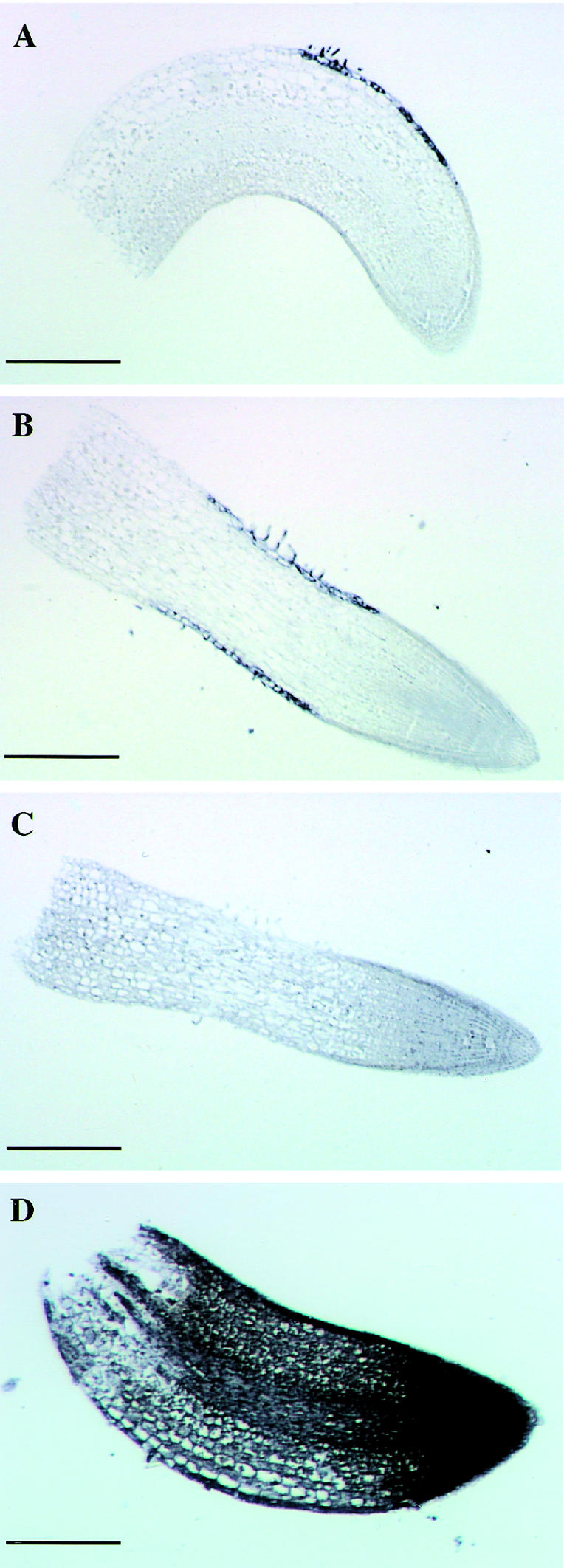
Figure 4.

Localization of LeExt1 transcripts in tomato seedling roots. A through D, Bright-field microscopy of root sections. Shown in A and B are sections hybridized with the LeExt1 antisense probe. The purple dye reflects LeExt1 mRNA. C, Section hybridized with the LeExt1 sense probe as a negative control. D, Section hybridized with an antisense Rpl2 probe as a positive control. Bar = 0.25 mm in A through C; bar = 0.125 mm in D.
