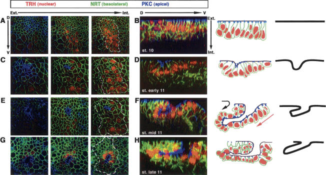
Figure 1.
Wild-type sequences of cell shape changes during tracheal invagination. Invagination of a tracheal placode is observed either using consecutive sections from the surface of the epithelium into the placode (A,C,E,G) or a single perpendicular section through the middle of the placode together with schematic representations (B,D,F,H). Here and in subsequent figures, anterior is to the left and dorsal to the top for the consecutive sections. For the perpendicular sections, dorsal is to the left and external to the top. The position of the tracheal placode is marked with a white dashed line. Tracheal cells are labeled using an anti-trachealess antibody (TRH). Anti-Neurotactin (NRT) labels the basolateral and basal sides of all epithelial cells, while PKC labels their apical side. In the schematic diagrams, the dark line delineates the apical surfaces of the cells. (A,B) At stage 10 before invagination, cells form a flat epithelium. (C,D) At the onset of invagination at early stage 11, a small group of cells has reduced its apical perimeter, and the epithelium begins to bend. Note that the most apical region of those cells lies in a deeper position than the neighboring ones. (E–H) As invagination proceeds during mid- and late stage 11, the apical surface of the invaginating cells is found in an even deeper position. Cells of the dorsal side of the placode have rotated completely around their axis and are found inside the embryo (black arrow), while ventral cells gradually slide beneath (red arrow), both movements leading to the formation of a finger-like structure.

