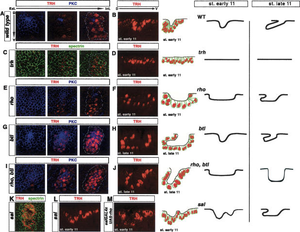
Figure 2.
Genetic control of cell shape changes during tracheal invagination. Consecutive confocal sections (A,C,E,G,I) and single perpendicular sections with schematic representation of the apical position of cells at early (B,D,F,L,M) and late (H,J) stage 11 in different mutant backgrounds. Here and in Figure 5, anti-α spectrin is used to label all epithelial cell membranes. (A,B) In wild-type embryos, invagination is initiated with the apical constriction of a small, spatially restricted, group of cells and curving of the epithelial layer. (C,D) In trh mutants, no sign of apical constriction is detected, and the epithelium remains flat. (E,F) In rho mutants, initiation of apical constriction within a few cells is not observed. Instead, aberrant invagination leads to the formation of a large cavity in the tracheal placode. By the end of stage 11, an abnormal finger-like structure is observed. (G,H) btl (FGFR−) mutants exhibit localized apical constriction and finger-like formation at the onset of invagination. Nevertheless, the finger does not extend further during stage 11. (I,J) Initiation of invagination in rho, btl placodes is as in rho mutants. However, at late stage 11, a finger-like structure is never observed. (K,L) In sal mutants, invagination is initiated at two positions and ultimately gives rise to an abnormal finger-like structure at late stage 11. (M) Similarly, overexpression of rhomboid driven by salGAL4 in the dorsal half of the placode (see Fig. 3) leads to formation of a double arch at the early stage 11 placode.

