Figure 3.
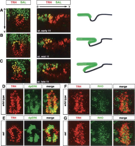
sal down-regulates EGFR activity in the dorsal tracheal cells. (A–C) The tracheal placode is divided into a dorsal, spalt (SAL)-positive domain, and a ventral, SAL-negative one. (A) The invagination point is located at the border between these two domains. (B,C) SAL-positive cells contribute to the formation of the roof of the extending finger. In the schematic diagram, black lines represent the apical surfaces of the tracheal cells, while green represents the domain of SAL expression. (D,E) dpERK is detected at higher levels in the ventral half of the wild-type placode, while the opposite is observed in sal mutants. (F,G) Rhomboid accumulation is higher on the ventral side of wild-type placodes (mean signal amplitude of the ventral side of 93.76 [SD 18.14]; cf. 58.65 [SD 16.69] of the dorsal side). In contrast, Rhomboid is more evenly distributed in sal mutants (mean signal amplitude of the ventral side of 122.17 [SD 24.96]; cf. 115.80 [SD 34.04] of the dorsal side).

