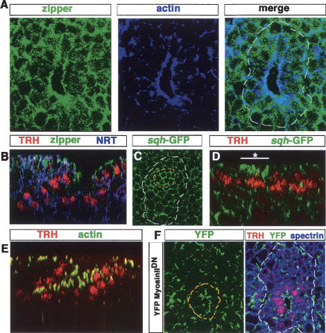
Figure 4.
Actin and myosin II distribution in tracheal cells. (A) Myosin II, as detected with an anti-zipper antibody, is specifically localized at the site of invagination. (B) Myosin II and actin colocalize at the invagination point. (C) Restricted distribution of zipper is seen in a perpendicular section. (D) Accumulation of Myosin II is detected at the onset of invagination in the cells initiating apical constriction (white line with an asterisk), as visualized by a GFP-tagged form of Myosin II light chain (sqh-GFP). (E) In contrast to the restricted zipper distribution, actin is strongly enriched apically in all tracheal cells during invagination. (F) A non-actin-binding YFP-Myosin IIDN accumulates irregularly at the invagination cells (outlined in yellow) forming noncontinuous apical patches. This mutated version of Myosin II construct is expressed using the GAL4 driver 69B, and its distribution is followed using an anti-GFP antibody.

