
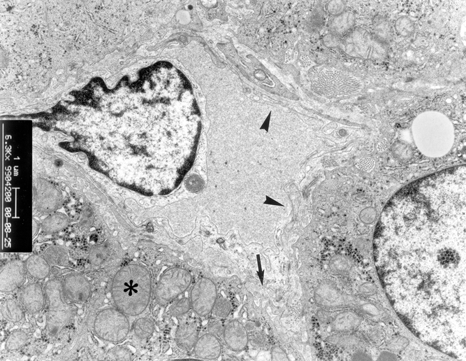
Figure 10. Electron microscopy examination of liver graft in patients in group 2. Slight mitochondrial swelling in hepatocyte was found (*). There were microvilli in the space of Disse (arrow). The sinusoidal lining cells were intact (arrowhead) (×6,300).