
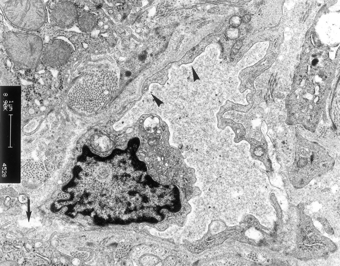
Figure 11. Electron microscopy examination of liver graft in patients in group 3. There were microvilli in the space of Disse (arrow). The sinusoidal lining cells were intact (arrowhead) (×6,300).
Official websites use .gov
A
.gov website belongs to an official
government organization in the United States.
Secure .gov websites use HTTPS
A lock (
) or https:// means you've safely
connected to the .gov website. Share sensitive
information only on official, secure websites.
Figure 11. Electron microscopy examination of liver graft in patients in group 3. There were microvilli in the space of Disse (arrow). The sinusoidal lining cells were intact (arrowhead) (×6,300).