
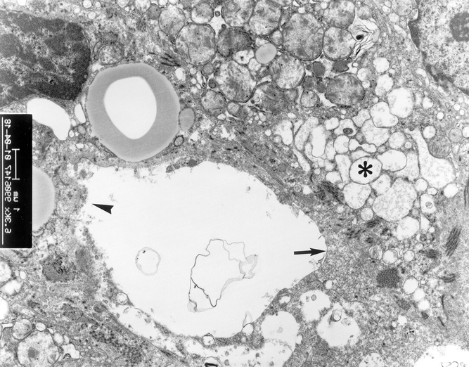
Figure 9. Electron microscopy examination of liver grafts in patients in group 1. Extensive swelling of hepatocyte mitochondria (*) and collapse of space of Disse (arrow) were found. Irregular large gap of sinusoidal lining cells was present (arrowhead) (×6,300).