
Figure 3.
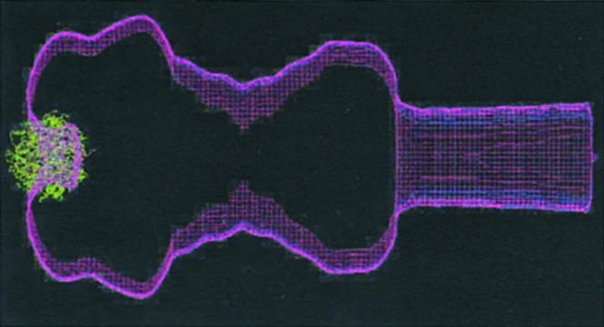
Docking of a Spa47 ATPase F1-like model (green) to the outline of the Shigella NC (purple). Spa47 was aligned to the sequences of α- and β- subunits of the F1-ATPase. Six different Spa47 monomer 3D models were generated and assembled by using whatif and the F1-coordinates (70). An outline of our NC reconstruction (10) was made by using spider and empirically scaled to the ATPase model by using the molecular dimensions of the complex measured in electron microscopy images (±15% accuracy). The ATPase was docked to the NC outline with ATP-binding sites facing the bacterial cytoplasm by using whatif.