Figure 5.
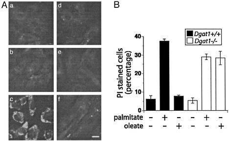
Lipotoxicity in Dgat1−/− cells. (A) Wild-type (Aa–Ac) and Dgat1−/− (Ad–Af) mouse embryonic fibroblasts were incubated with media alone (Aa and Ad) or media supplemented with 1 mM palmitate (Ab and Ae) or 1 mM oleate (Ac and Af) for 12 h. Triglyceride accumulation was detected by Nile red staining. Bar = 20 μm. (B) Wild-type and Dgat1−/− fibroblasts were supplemented with 1 mM palmitate or 1 mM oleate for 46 h. Cell death was assessed by PI staining and flow cytometry. For each sample, fluorescence of 104 cells was assessed, and the percentage of cells with PI fluorescence was determined. The bar graph displays the median percentage of PI-positive cells from three independent measurements ± SE.

