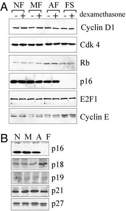
Figure 2.
Expression of cell-cycle regulatory proteins during FS progression. (A) Cells from each stage (clones as indicated in Fig. 1) were cultured in the absence (−) or presence (+) of 100 nM dexamethasone for 48 h. Whole-cell extracts were probed with antibodies against cyclin D1, cdk4, pRb, p16, E2F1, and cyclin E (antibodies were used as described in Materials and Methods). Each immunoblot is representative of at least three independent experiments. (B) Intracellular levels of various cdk inhibitors during FS progression. A second set of clones, different from the set used above, and representative of each stage of FS progression (NF non-BPV, MF 39614, AF BPV7, and FS BPV22, represented by N, M, A, and F, respectively) were used in this experiment. Each blot was reprobed for β-tubulin as a control for equal loading (data not shown). Note that the apparent increase of p18 expression in FS cells was insignificant after correction with tubulin levels.

