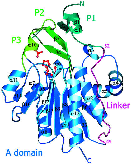
Figure 1.
Ribbon representation of the EcHsp31 monomer. The A domain is blue, the P domain is green (P1–P3 segments are in progressively lighter shades of green), and the linker region is purple. The catalytic triad is shown as a red ball-and-stick model (28).

