Figure 1.
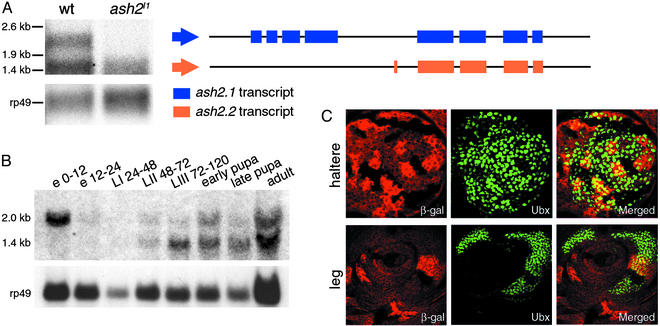
(A–C) Molecular characterization of WT and mutant ash2 mRNA. (A) Northern blot analysis of poly(A) RNA extracted from WT and ash2I1 homozygous third instar larvae. Structure of the 2- (ash2.1) and 1.4-kb (ash2.2) transcripts after performing rapid amplification of cDNA ends is shown. (B) Developmental Northern blot of ash2 expression. In A and B, rp49 was used as a loading control. e, embryo; L, larval stages; numbering indicates hours after egg laying. (C) Down-regulation of UBX protein accumulation in ash2I1 mutant clones generated in haltere and leg imaginal discs by FLP-FRT-induced mitotic recombination. (Left) Staining with anti-β-galactosidase antibody:WT cells (bright red), heterozygous cells (red), and homozygous ash2I1 mutant cells (lack of red staining). (Center) Staining with anti-UBX antibody (green). (Right) Merged images.

