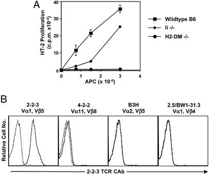
Figure 1.
Characterization of the 2-2-3 hybridoma and 2-2-3 clonotypic Ab. (A) The 2-2-3 T cell hybridoma recognizes an I-Ab-associated peptide. 2-2-3 hybridoma T cells (1 × 10⁵) were stimulated with increasing numbers of splenocytes from the indicated mice, and IL-2 in the supernatant was assayed by using the HT-2 cell line. (B) The 2-2-3 clonotypic Ab recognizes only 2-2-3 T cells. The T cell hybridomas shown were stained with 2-2-3 clonotypic Ab followed by FITC-conjugated goat anti-mouse IgG (solid line). The dashed line shows staining with secondary Ab alone.

