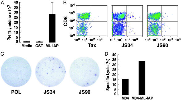
Figure 3.
ML-IAP-specific T cells were associated with tumor destruction. (A) Proliferative responses of CD4+ T cells derived from the calf metastasis. (B) JS34- and JS90-tetramer-stained CD8+ T cells from the colon metastasis. CD8+ tetramer+ cells were as follows: JS34, 1.01%; JS90, 0.86%; and Tax, 0.38%. (C) JS34 and JS90 peptides elicited IFN-γ production from CD8+ T cells in the colon metastasis. Spots shown were (no. of replicate wells) as follows: Pol (0, 3); JS34 (20, 4, 10); and JS90 (3, 7, 6). No reactivity was detected with autologous peripheral blood mononuclear cells alone (data not shown). (D) T cells from the calf metastasis showed ML-IAP-specific cytotoxicity (effector-to-target cell ratio of 30:1).

