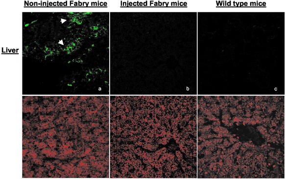
Figure 3.
Immunostaining for Gb3 in mouse liver after administration of rAAV-CAG-hAGA. Tissue specimens from noninjected Fabry mice, injected Fabry mice, and wild-type mice were analyzed at 4 weeks postinjection. Samples were examined by confocal microscopy (×40). (Upper) Gb3 staining with FITC-conjugated anti-Gb3 antibody. (Lower) Nuclear staining in the same sections with propidium iodide. There is intense Gb3 fluorescence in noninjected Fabry mice compared with that of injected Fabry or wild-type mice.

