
Figure 1.
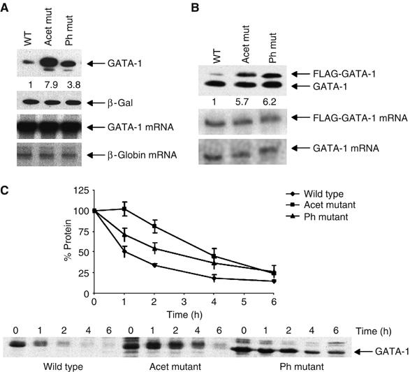
Mutation of phosphorylation and acetylation sites increases GATA-1 stability. (A) Upper panels: Western blot showing GATA-1 levels in Cos 7 cells, transfected with constructs to express wild-type GATA-1 (Wt), phosphorylation (Ph) or acetylation (Acet) mutants. β-Galactosidase is the cotransfection control. Lower panels: RNase protection assay of the corresponding GATA-1 mRNAs; the human β-globin gene is the cotransfection control. (B) Upper panel: Western blot showing the levels of endogenous and FLAG-tagged GATA-1 protein in BM-SCF cells stably infected with retroviruses to express FLAG-tagged versions of GATA-1. Lower panels: RNase protection assay of the FLAG-tagged and endogenous GATA-1 mRNAs. (C) Pulse–chase experiment to determine the half-life of GATA-1. The levels of wild type (⧫), acetylation (Acet ▪) and phosphorylation (Ph ▴) mutants in Cos 7 cells are shown. A value of 100% was given to the amount of protein at time zero; the per cent of protein remaining was calculated relative to this. The plot shows the average of three experiments; error bars show standard error.