
Figure 5.
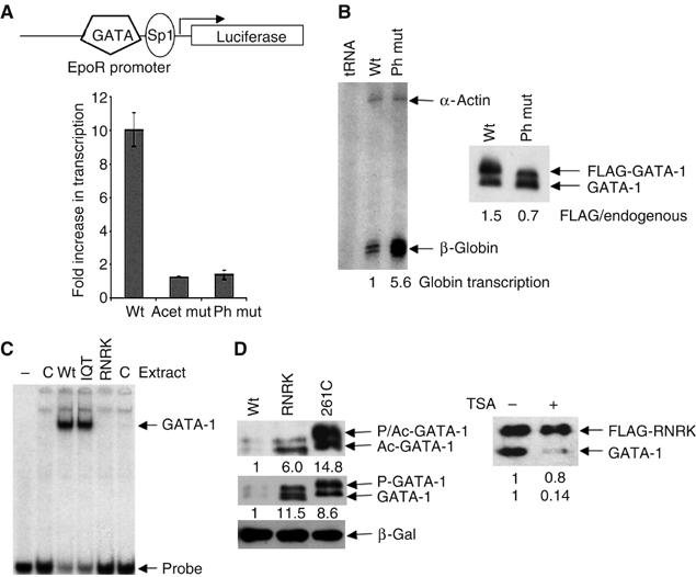
Preferential degradation of transcriptionally active GATA-1. (A) Upper: The reporter construct pGL3EpoR. Lower: Inhibition of phosphorylation increases GATA-1-dependent transcription. NIH3T3 cells, transfected with the constructs shown, were treated with or without the MEK inhibitor U0126. The transcription level in the presence of U0126 divided by that in its absence is plotted. The average of four experiments is shown. (B) The phosphorylation mutant is more transcriptionally active than wild-type GATA-1. Left panel: S1 mapping of β-globin gene transcription in BM-SCF cells that express FLAG-tagged wild-type (wt) or phosphorylation mutant (ph mut) GATA-1. Quantification of β-globin transcription, normalised to α-actin, is shown beneath the gel. Right panel: Western blot showing the level of the FLAG-tagged wild-type or phosphorylation mutant protein and endogenous GATA-1. For this experiment, cell lines were chosen where mutant and wild-type GATA-1 are expressed at similar levels. (C) The RNRK mutant does not bind DNA. Equivalent amounts of whole-cell extract prepared from mock-transfected Cos 7 cells (C) or cells that had been transfected with expression vectors for wild-type (wt) or mutant GATA-1 proteins (IQT and RNRK) were used in a gel mobility shift assay under the conditions described (Boyes et al, 1998). Control experiments confirmed that transfection efficiency was equivalent. (D) The DNA binding mutants are more stable than wild-type GATA-1. Left panel: Western blot of extracts from Cos 7 cells transfected with wild-type GATA-1 and the DNA binding mutants, C261P and RNRK, probed with anti-GATA-1 antibody or anti-acetyl-GATA-1 antibody. β-Galactosidase is the cotransfection control. Right panel: Haemopoietic (BM-SCF) cells, expressing FLAG-tagged RNRK GATA-1, were treated with or without TSA. A Western blot probed with anti-GATA-1 antibody (N-6) is shown.