
Figure 2.
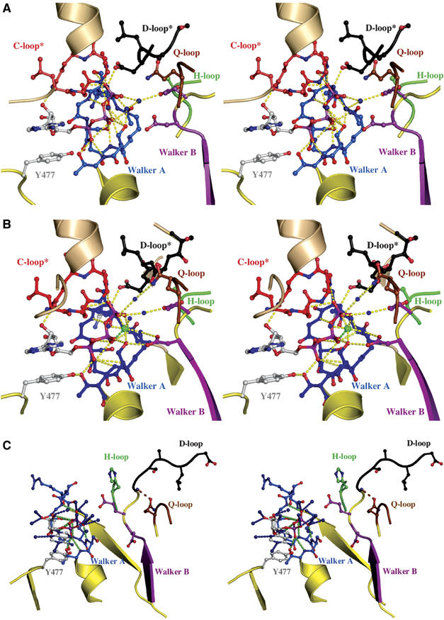
Nucleotide-binding sites. Stereoview of the ATP-binding (A) and ATP/Mg2+-binding(B) sites. Color-coding is identical to Figure 1. Direct and water-mediated protein–ATP interactions are highlighted in yellow. Water molecules are shown as blue spheres and Mg2+ as a green sphere. The interaction between D637 of the D-loop of the trans monomer and S504 of the Walker A motif of the cis monomer is indicated. ATP and amino acids involved in ligand interactions are shown in ball-and-stick representation. * indicates conserved motifs of the trans monomer participating in ATP coordination. (C) Stereoview of the ADP-binding site. ADP and residues involved in ligand interactions are shown in ball-and-stick representation, water molecules are blue spheres, protein–ADP interactions are highlighted in green and ADP–water interactions in blue. Color-coding is identical to Figure 1. The interaction between the side chain of Q550 and the amide backbone of T633 is highlighted by a dashed, brown line.