
Figure 5.
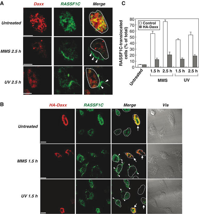
Daxx overexpression inhibits DNA damage-induced translocation of RASSF1C from the nucleus to the cytoplasm. (A) Endogenous RASSF1C translocation to the cytoplasm in response to DNA damage. HeLa cells that were left untreated or treated with MMS or UV were fixed at 2.5 h post-treatment and immunostained with anti-RASSF1C and anti-Daxx antibodies. White arrowheads indicate the accumulation of endogenous RASSF1C in the cytoplasm. White dots outline the nucleus. Scale bar, 5 μm. (B, C) Daxx inhibits RASSF1C translocation to the cytoplasm. (B) Single-cell immunohistochemical analysis. HeLa cells were transfected with pCMV-HA-Daxx and were left untreated or treated with MMS or UV. Cells were fixed at 1.5 h post-treatment and immunostained with anti-HA and anti-RASSF1C antibodies. White arrowheads indicate the accumulation of RASSF1C in the cytoplasm. White arrows indicate cells expressing HA-Daxx. White dots outline the nucleus. Scale bar, 5 μm. (C) Statistical analysis. The number of cells in (B) showing RASSF1C translocation to the cytoplasm at 1.5 and 2.5 h post-treatment was counted in 100 cells/sample and expressed as a percentage of the total cell number. Data shown are the mean percentage±s.e.m. of data obtained from at least three independent experiments.