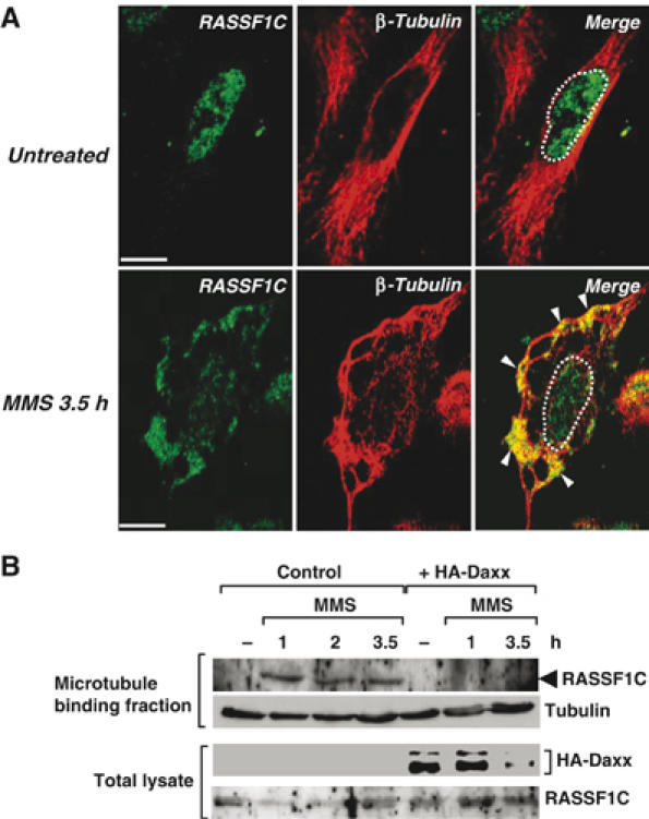
Figure 6.

Endogenous RASSF1C is translocated from the nucleus to the cytoplasmic microtubules in response to DNA damage. (A) HeLa cells were left untreated or treated with MMS and immunostained with anti-RASSF1C and anti-β-tubulin antibodies at 3.5 h post-treatment. White arrowheads indicate the accumulation of endogenous RASSF1C at the microtubules. White dots outline the nucleus. Scale bar, 5 μm. (B) Microtubule cosedimentation. HeLa cells transfected with pCMV-HA-Daxx or pCMV empty vector were left untreated or treated with MMS. The cell lysates were incubated with taxol to stabilize microtubules. The microtubule-binding fraction was isolated as described in Materials and methods and immunoblotted with anti-RASSF1C and anti-β-tubulin antibodies.
