
Figure 1.
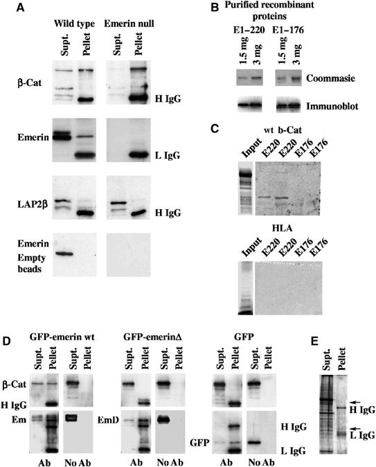
β-Catenin binds to emerin through a conserved APC homology domain. (A) β-Catenin was immunoprecipitated from cell extracts prepared from wt or emerin null fibroblasts. Immunoprecipitates were resolved on SDS–PAGE and immunoblotted with antibodies against total β-catenin (β-cat), emerin or LAP2β (top three panels). Alternatively, empty immunobeads were incubated with the same cell extracts and used for immunoblotting with anti-emerin antibody (bottom panel). The antibody used in each blot is indicated to the left-hand side of each panel, while the positions of IgG heavy or light chains (H IgG and L IgG, respectively) are indicated to the right-hand side of each blot. (B) Purified recombinant proteins E1–176 and E1–220 were resolved on SDS–PAGE, which were either stained with Coomassie Brilliant Blue (upper panels) or immunoblotted with anti-emerin antibody (lower panels). (C) 35S-met-labelled wt β-catenin or HLA were resolved on SDS–PAGE and subjected to autoradiography (input). Purified emerin E1–220 and E1–176 at 1.5 and 3 μg were resolved on SDS–PAGE and transferred to nitrocellulose. The filters were then overlayed with 35S-methionine-labelled proteins and subjected to autoradiography. In all panels, the first lane was loaded with 1.5 μg of recombinant protein, while the second lane was loaded with 3.0 μg. (D) β-Catenin and one of GFP-emerin, GFP-emerinΔ or GFP were coexpressed in HEK293 cells. Cell lysates were immunoprecipitated with anti-GFP antibody or empty immunobeads (indicated by Ab or no Ab, respectively, underneath each panel). Immunoprecipitates were immunoblotted with antibodies against β-catenin (upper panels) or GFP (lower panels). The positions of β-catenin (β-cat), GFP-emerin (em), GFP-emerinΔ (emΔ) or GFP are indicated at the left-hand side of each panel. The positions of IgG heavy chain (H IgG) and IgG light chain (L IgG) are indicated at the right-hand side of the final panel. (E) HEK293 cells that were co-transfected with GFP-emerin and β-catenin were immunoprecipitated with anti-GFP antibodies, resolved on SDS–PAGE and stained with ammoniacal silver. In all gels, Supt=material remaining in the supernatant. Pellet=material recovered in immunoprecipitates. Arrow indicates a band with expected mobility of GFP-emerin.