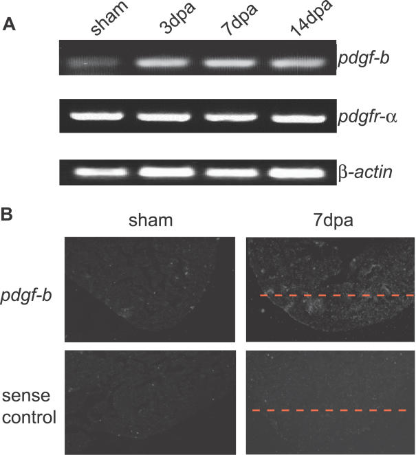
Figure 4. pdgf-b Expression Is Upregulated in Regenerating Zebrafish Hearts.
(A) RT-PCR analysis of pdgf-b and pdgfr-α. β -actin was used as a loading control. Expression of pdgf-b began to increase at 3 dpa and lasted until 14 dpa during zebrafish heart regeneration. pdgfr-α was expressed in both sham-operated and regenerating hearts; its expression level does not change.
(B) In situ hybridization using a radioactive antisense probe showing pdgf-b expression in sham-operated and regenerating hearts at 7 dpa. Expression of pdgf-b was localized to the wound site. A radioactive sense probe was used as a negative control. The dashed red line marks the amputation plane.

