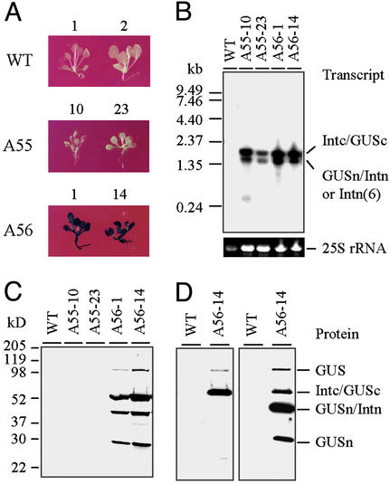
Figure 3.
Examination of in vivo assembly of the divided β-glucuronidase fragments in 2-wk-old Arabidopsis seedlings. (A) GUS-staining assay. (B) RNA- filter hybridization assay. Total RNA (≈6 μg) was loaded in each lane and probed with 32P-labeled GUS-coding sequence. The 25S rRNA was used as a loading control in each lane. (C and D) Protein immunoblot assays. Total soluble protein (15 μg) was loaded in each lane. (C) Protein was probed with Penta-His Ab. (D) The protein was probed with anti-His(C Term)-HRP Ab at first (Left). After stripping, it was reprobed with Penta-His Ab (Right). For each assay, results obtained from two individual events of A55 and A56 transformations are shown. WT plants were used as controls.

