Figure 5.

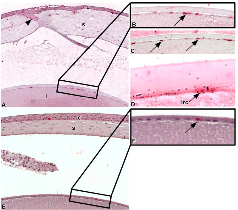
Wounding stimulates the central lens epithelial cells to proliferate. Scrape wounds were created on the lenses of mice that were labeled with an in utero/post-natal protocol and chased for 18.5 weeks. Twenty-four hours post-wounding, BrdU was injected intraperitoneally to tag proliferating cells. These mice were sacrificed 24 hours later (A) and paraffin sections were processed for BrdU immunohistochemistry and autoradiography. Arrowheads demarcate wound area. Note the increased number of BrdU labeled cells in corneal (c) and lens (l) epithelia. A portion of the central zone of the lens epithelium (area in the rectangle) containing BrdU labeled cells (arrow) is shown in higher magnification (B). (C) Paraffin section of a portion of the central zone of the lens epithelium from another mouse 24-hrs post-wounding showing numerous flattened lens epithelial cells stained with BrdU (arrows). (D) A central lens epithelial cell is double-labeled, containing both 3H-TdR and BrdU. This indicates that LRCs (lrc) are healthy and can divide following wounding. (E) A portion of a non-wounded, resting cornea (c) and lens (l) that were labeled with a single intraperitioneal injection of BrdU and sacrificed 24 hours later. A portion of the central zone of the lens epithelium (area in the rectangle) containing a single BrdU-labeled cell (arrow) is shown in higher magnification (F).
