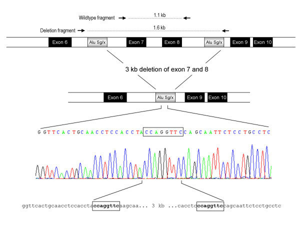
Figure 2.
Schematic representation of the mutation deleting exon 7 and 8 of the LDLR gene. The locations of the primers used for the diagnostic duplex PCR is shown. Deletion breakpoints are flanked by Alu Sg/x elements (grey boxes). The box in the sequence data represents nucleotides present in each end of the breakpoint of the reference sequence shown below the sequence trace.

