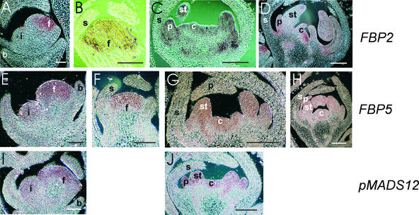
Figure 4.
In Situ Localization of FBP2, FBP5, and pMADS12 Transcripts in Wild-Type Petunia.
Longitudinal sections were hybridized to digoxigenin-labeled antisense RNA fragments of FBP2 ([A] to [D]), FBP5 ([E] to [H]), and pMADS12 ([I] and [J]).
(A) FBP2 expression in a very young floral meristem. No signal is obtained in the inflorescence meristem and bracts.
(B) FBP2 expression in the central part of an early-stage floral bud. No signal is present in the developing sepals.
(C) Young floral bud with developing sepal, petal, and stamen primordia. The carpel primordia just arise. FBP2 mRNA can be detected in the three inner floral whorls.
(D) FBP2 expression in a slightly later stage floral bud than that shown in (C).
(E) Same stage meristems as in (A) hybridized to an antisense FBP5 probe. In contrast to FBP2, FBP5 also is expressed in the inflorescence meristem.
(F) FBP5 expression in an early floral bud showing a pattern similar to that obtained for FBP2 (B).
(G) and (H) FBP5 expression in the inner three floral whorls. Stages are comparable to those shown in (C) and (D), respectively.
(I) Expression of pMADS12 in the inflorescence and floral meristem. The expression pattern resembles the expression of FBP5 at this stage (E).
(J) pMADS12 expression in a same stage floral bud as that shown in (C) and (G).
b, bract; c, carpel; f, floral meristem; i, inflorescence meristem; p, petal; s, sepal; st, stamen. Bars in (A), (B), (E), (F), and (I) = 200 μm; bars in (C), (D), (G), (H), and (J) = 500 μm.

