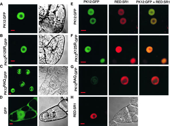
Figure 2.
Subcellular Localization of PK12 and SR1 Fusion Proteins.
Confocal optical sections are shown of viable BY-2 cells that overexpress either PK12 or SR1 proteins fused to GFP and dsRED, respectively, or cells that overexpress both PK12:GFP and RED:SR1. The panels at right show color overlays. The fluorescence images displaying either GFP or dsRED signal are shown together with the corresponding Nomarski images. Bars = 10 μm.
(A) PK12:GFP.
(B) PK12K125R:GFP.
(C) PK12RAQ:GFP shown in cells exhibiting nuclear ring structure.
(D) GFP.
(E) PK12:GFP and RED:SR1.
(F) PK12K125R:GFP and RED:SR1.
(G) PK12RAQ:GFP shown in cells displaying nuclear ring structure and RED:SR1.
(H) RED:SR1.

