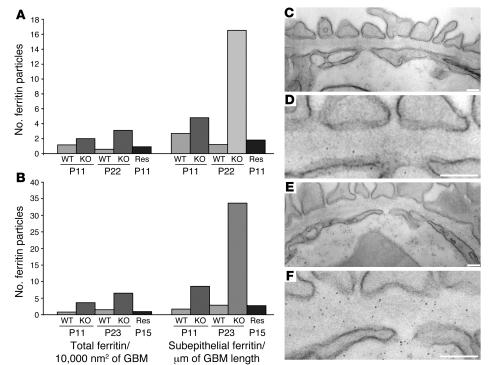
Figure 5. Increased GBM permeability to ferritin inLamb2–/– mice.
Ferritin was detected in the GBM either 1 (A) or 2 (B) hour after a single intravenous injection into control/mutant littermate pairs, at the indicated ages. Ferritin particles were counted in the total surface area of the GBM (expressed as number of ferritin particles/10,000 nm2) or only at the subepithelial (podocyte) aspect (expressed as number of ferritin particles/μm of GBM length). There was an increase in total and in subepithelial ferritin at 1 hour in all Lamb2–/– mice compared with control or non-nephrotic, rescued Lamb2–/– mice carrying the NEPH-B2 transgene (Res). The increase was more remarkable after 2 hours in the older mice. (C–F) Representative electron micrographs showing ferritin particles in the GBM of normal (C and D) and Lamb2–/– (E and F) mice at P11. Note the increased ferritin in the mutant GBM despite the normal FP architecture. D and F are higher magnifications of C and E, respectively. Scale bars: 125 nm.

