
Fig. 3.
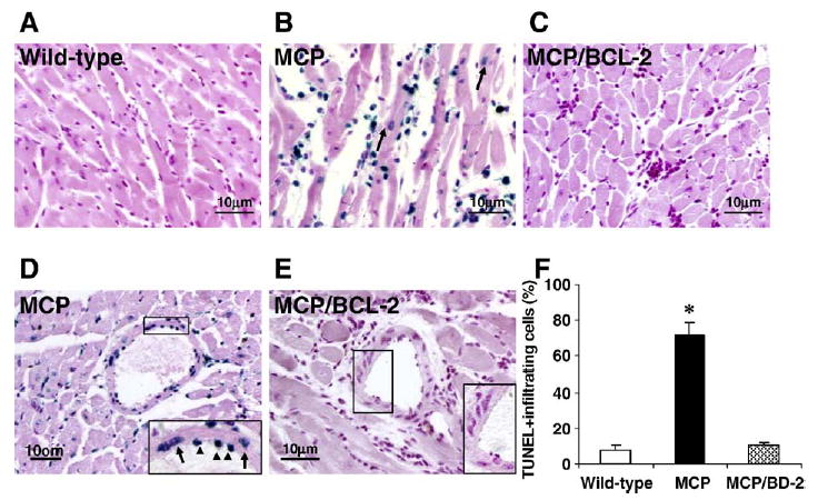
Bcl-2 expression in monocytes prevents apoptotic cell death in the hearts of MCP mice. (A–E) Representative photomicrographs demonstrating TUNEL staining of heart sections from 6-month-old wild-type, MCP and MCP/Bcl-2 mice. Blue staining indicates TUNEL-positive cells and the TUNEL-positive cardiac myocytes are indicated by arrows in B. The insert in D shows TUNEL-positive vascular endothelial (arrowheads) and smooth muscle cells (arrows) in the heart of MCP mouse. The insert in E shows no TUNEL-positive vascular cells in the heart of MCP/Bcl-2 mouse. (F) Histogram showing the quantitative analysis of TUNEL-positive infiltrating cells in the hearts of mice at 6 months of age. n =5 per group. *P <0.001 versus wild-type mice and MCP/Bcl-2 mice.