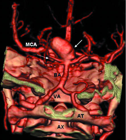
Fig 2.
Volume rendered computed tomography angiogram, viewed from posterior to anterior, showing a 20 mm diameter aneurysm (solid arrow) at tip of basilar artery (BA) anda6mm diameter aneurysm (dashed arrow) on the left posterior communicating artery. MCA=left middle cerebral artery; VA=left vertebral artery; AT=atlas (C1); AX=odontoid process of axis (C2)

