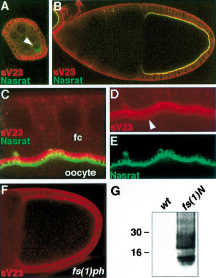
Figure 3.
Nasrat and Polehole are required for cross-linking of sV23 product. (A–E) Egg chambers expressing the Nasrat–Flu construct stained with anti-Flu (green) and anti-sV23 (red). (A) Stage 5 egg chamber; Nasrat and sV23 show cytoplasmic accumulation in the oocyte (arrowhead) and follicle cells, respectively. (B) Stage 10 egg chamber; Nasrat and sV23 mark the intercellular space between the oocyte and the follicle cells. (C–E) Close-up view of the stage 10 follicle cortex. Nasrat and sV23 outline the microvilli formed by the oocyte and follicle cells (fc), respectively. We also detect low levels of sV23 on the oocyte surface (arrowhead, D). (F) sV23 staining in a mutant fs(1)phK646 egg chamber that lacks both Nasrat and Polehole at the oocyte periphery; sV23 appears to accumulate normally. (G) Western blot analysis with sV23 antibody of protein extracts from embryos laid by wild-type and fs(1)N14 mutant mothers; note the presence of soluble sV23 species in the vitelline membrane of mutant embryos.

