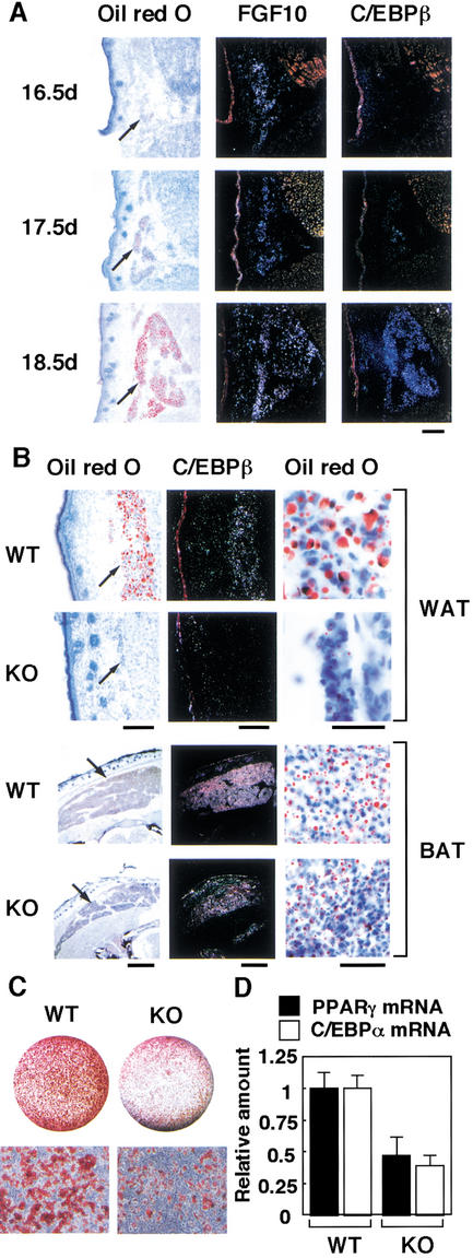
Figure 5.
Expression of FGF10 genes in WAT during mouse embryogenesis, histological analysis of WAT and BAT of FGF10 knockout neonates, and the ability of EF to differentiate into adipocytes. (A) Expression of FGF10 and C/EBPβ genes in subcutaneous WAT of wild-type mouse embryos at E16.5, E17.5, and E18.5. Sections were stained with oil red O or subjected to in situ hybridization with 35S-labeled FGF10 or C/EBPβ antisense RNA probes. Scale bar, 100 μm. Arrows indicate subcutaneous WAT. (B) Subcutaneous WAT and interscapular BAT of FGF10 knockout (KO) mice and wild-type (WT) littermates (within 10 min after birth) were stained with oil red O or subjected to in situ hybridization with a 35S-labeled C/EBPβ antisense RNA probe. Scale bars, 100 μm. Macroscopic (left panels) and microscopic (right panels) views of oil red O staining are shown. Arrows indicate WAT and BAT. At least three neonates of each genotype were examined, and representative data are shown. (C,D) EF obtained form FGF10 knockout mice or wild-type littermates were incubated in the inducers of differentiation for 8 d, then the cells were stained with oil red O (C), or the abundance of C/EBPα or PPARγ mRNA was determined by real-time PCR (D). (C, upper panels) Macroscopic and (C, lower panels) microscopic views of oil red O staining. Data in C are representative of three experiments and in D are means ± SEM of values from six experiments.

