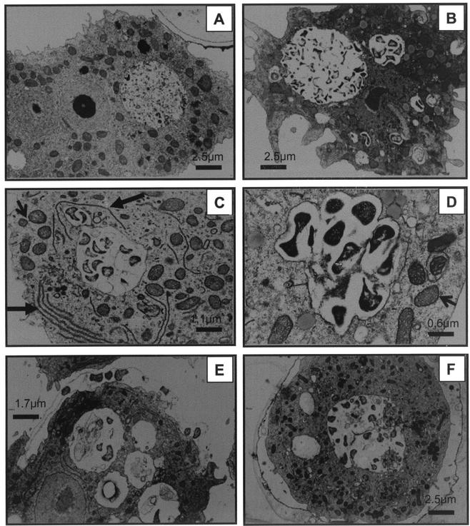
FIG. 4.
Electron microscopy analysis. (A) A. castellanii trophozoite without intracellular F. tularensis (day 0). (B) A. castellanii trophozoite with Francisella-filled vacuoles (day 9). (C and D) Recruitment of mitochondria (short arrows) and rough endoplasmic reticulum (long arrows) to the vacuole containing bacteria. (E) A. castellanii trophozoite undergoing encystation with F. tularensis cells lined up between the two layers of the emerging double wall (day 16). (F) A. castellanii cyst containing F. tularensis on the inside of the double wall (day 16).

