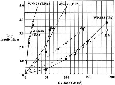
FIG. 1.
Summary of spore inactivation by 254-nm UV. B. subtilis biodosimetry strains WN333 (circles) and WN626 (triangles) at EPA (open symbols) or at UA (filled symbols). For comparison, the UV inactivation kinetics from Table 1 are plotted for spores of E. intestinalis (solid squares), E. cuniculi (hatched squares), and E. hellem (open squares). The data points shown for WN333 at UA are averages of three separate determinations, which varied by ±5%.

