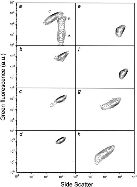
FIG. 4.
Flow cytometric characterization of E. coli NM522(pML1) with regard to SSC and DiBAC4(3)-derived fluorescence after chemical and heat treatment and comparison to the bacterial populations (a) detected 20 min after lysis induction: nonlysed, polarized cells (A); nonlysed, depolarized cells (B); and bacterial ghosts (C). (Note: panel a corresponds to Fig. 3, +20 min.) Log-phase growing cells were treated with heat (b), ethanol (c), 2,4-dinitrophenol (d), pore-forming colicin E1 (e), formaldehyde (f), or ampicillin (g). An aliquot of growing cells was subjected to a procedure developed by Lam and colleagues (13) for the preparation of outer membrane fractions of H. influenzae (h). The cytometric analysis data, which are derived from 104 bacterial particles (b to h) or the particles within 0.1 μl of the lysing culture (a), are presented as contour plots (SSC versus green fluorescence in arbitrary units [a.u.], both on a logarithmic scale) excluding debris and the background signal.

