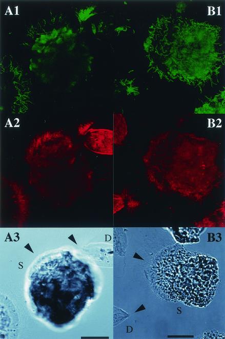
FIG. 4.
Detection by in situ hybridization of spirochetes attached to the gut protists Devescovina sp. and Stephanonympha sp. (A1 and B1) Epifluorescent images with probes specific for phylotypes NkS-Dev1 (Dev1-486) and NkS-Dev14 (Dev14-486 and Dev14-Tp654), respectively. (A2 and B2) Epifluorescent images with the eubacterial universal probe (EUBAC). Note that rod-shaped (nonspiral) bacteria are also attached to the cells of Devescovina sp. (A3 and B3) Differential interference contrast micrographs. The protist species are labeled as D (Devescovina sp.) and S (Stephanonympha sp.). Bars, 20 μm. The arrowheads indicate the anterior parts of the protist cells.

