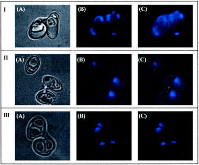
FIG. 3.
Microscopy of stained Z. bailii cells. (A) Bright field; (B) blue fluorescence; (C) overlap of bright field and blue fluorescence. (Section I) Conjugated cells stained with calcofluor white; (section II) conjugated but not sporulated cells stained with DAPI; (section III) sporulated cells stained with DAPI. The fluorescence of calcofluor white and DAPI is blue.

