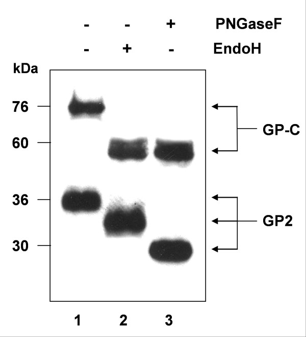
Figure 4.
Glycosidase sensitivity of cell surface transported Lassa virus glycoprotein. At 48 h post-transfection, Vero cells expressing wild-type Lassa virus glycoprotein were surface biotinylated and immunoprecipitated as described under Fig. 3. Precipitated samples were either left untreated (lane 1), digested with Endo H (lane 2) or PNGase F (lane 3). The samples were separated by SDS-PAGE with subsequent immunoblotting. Biotinylated proteins were detected using streptavidin coupled to horse radish peroxidase and chemiluminescence.

