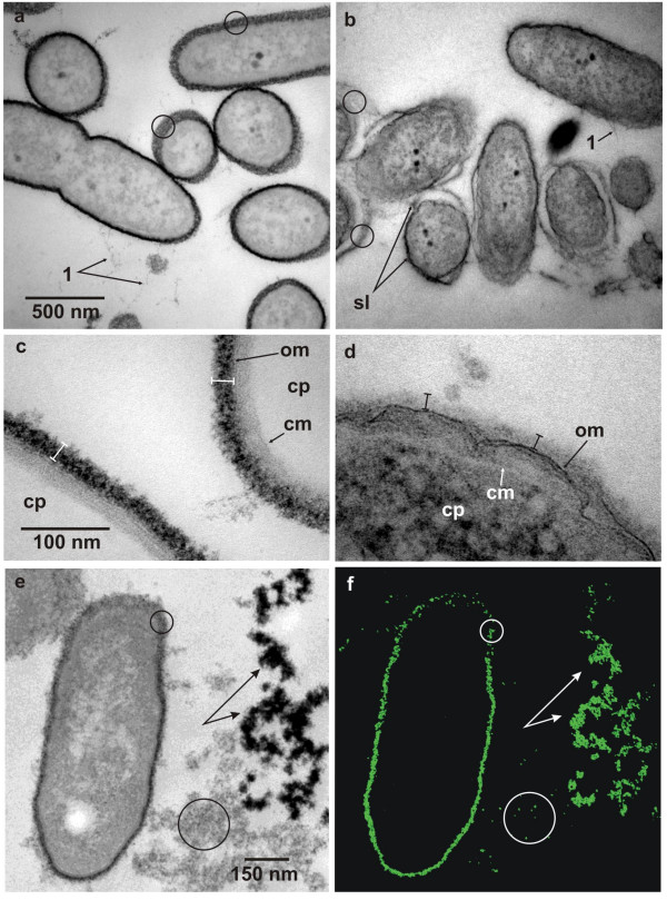
Figure 2.

Ultrathin sections of pre-embedding thorium dioxide label of cellular slime layers of Pseudomonas aeruginosa. (a) The outer surface of the cells is intensely stained with thorium dioxide colloids and from obliquely cut cells or cells cut non-equatorially this layer appears granular and non-fibrillar in substructure (circles). Fine fibrillar matter emanates from the cell surface (1) and shows a fine granular substructure. (b) Acidic mucopolysaccharide staining with AlcianBlue™. An electron dense slime layer (sl) envelopes the bacterial cells. This matrix irregularly contours the cell, either directly attaching the surface or running at varying distances. Obliquely cut layers show fibrillar rather than granular substructures (circles). (c) Detailled view of thorium dioxide label of the outer surface layer reveals a high density and granularity of slime layer (spacer bars), outer (om) and cytoplasmic membranes (cm) outline the periplasm, the cytoplasm (cp) is unstained and appears electron translucent. (d) Treatment of outer surface layer with RRL reveals a weakly greyish stained matrix (spacer bars). The outer (om) and cytoplasmic membranes (cm) are clearly visible as indicated, contouring the cytoplasm (cp). (e, f) Elastic bright field view (e) of a bacterial cell and adjacent EPS and the corresponding Th-elemental map (f) (section thickness: 35 nm). Because of the oblique orientation of the cell wall Th-densities in this area (small circles) are relatively low compared to the perpendicularly oriented parts. Large circles enclose EPS with low Th-densities and branched arrows indicate high Th-densities of EPS clusters with high negative intrinsic charges. Bars in (a, c, e) are valid for (b, d, f).
