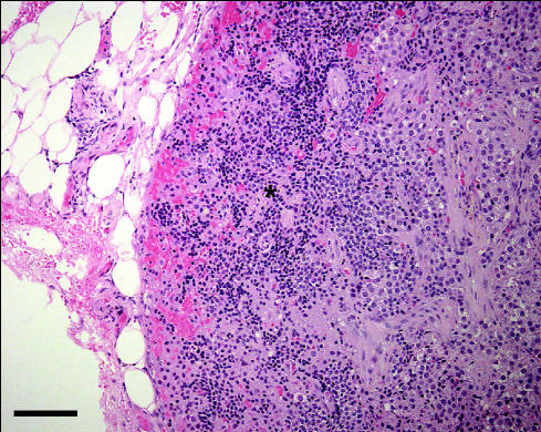
Figure 2.
A higher magni.cation image of the mass as in Figure 1. The small cells with the small, dark staining nuclei at the edge of the mass (*) were interpreted to be normal parathyroid gland cells that had been displaced and compressed by the expanding adenoma. Hematoxylin and eosin stain; bar = 10 μm.

