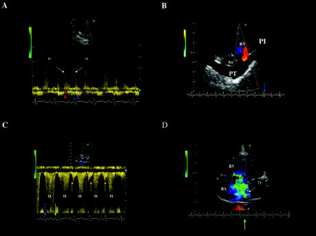
Figure 1. Doppler echocardiograms from the dog with severe pulmonary arterial hypertension.
A. The continuous-wave Doppler tracing of the right ventricular outflow tract obtained from the right parasternal short axis position showed diastolic pulmonary insufficiency (positive waves, PI) beginning after the T wave (red arrow) and ending during the isovolumic contraction period (blue arrow).
B. The prolongation of pulmonary insufficiency (PI) during isovolumic contraction (blue arrow) was confirmed by color-flow Doppler from the right parasternal short axis position. Blood flow (red color) is directed toward the transducer into the right ventricular outflow tract.
C. Continuous-wave Doppler examination performed from the left apical 4-chamber view identifying a marked tricuspid insufficiency (negative waves, TI) beginning during the isovolumic contraction phase (blue arrow), continuing during systole and isovolumic relaxation, and ending in the middle of P wave (green arrow). A late diastolic negative signal occurring after the P wave onset can also be seen (yellow arrows). This may arise from diastolic tricuspid regurgitation or from reversal of flow (A reversal) in the caudal vena cava.
D. Prolongation of the tricuspid insufficiency (TI) during early electrical diastole was confirmed using color-flow Doppler mode, frame taken between the end of T wave and the beginning of P wave (green arrow). The large turbulent jet of TI extends from the right ventricle to the dorsal wall of the severely enlarged right atrium.
RA — Right atrium; RV — Right ventricle; Ao — Aorta; PT — Pulmonary trunk

