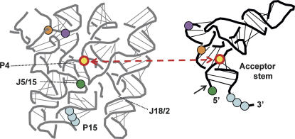
FIGURE 3.
Three-dimensional representation of Type B RNase P and tRNA adapted from Kazantsev et al. (2005, © National Academy of Sciences, USA). Colored dots in blue, green, orange, and purple represent interactions previously identified between ribozyme and substrate. Yellow dots represent positions involved in the XLU69 cross-link obtained here using E. coli (Type A) RNase P. A black arrow indicates the site of pre-tRNA cleavage.

