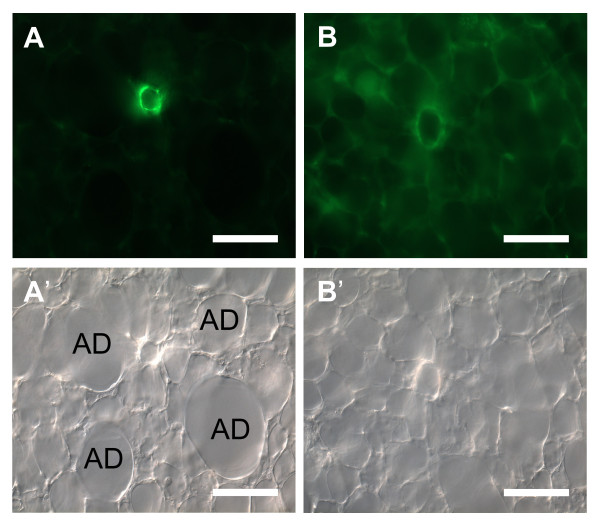
Figure 3.
Difference between arteries and veins in PCLS. Arteries in cross sections (A, A') can be easily distinguished from veins (B, B') by their accompanying alveolar ducts (AD) in the bright field mode here shown in differential interference contrast (A', B'). The muscular coat of arteries is thicker than in veins as revealed by α SMA-immunohistochemistry (A, B). Bars = 100 μm.

