
Fig. 1.
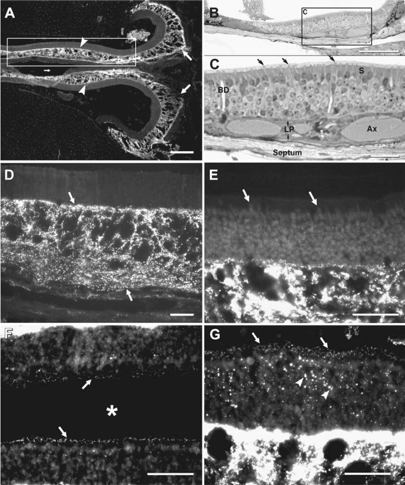
Immunofluorescence labeling of Cx43 in adult mouse OE. (A) Low magnification micrograph of a midline horizontal section through the posterior pole of the OE abutting the osseous nasal septum medially (small arrow) and the cribriform plate posteriorly (right, large arrows). On both left and right sides, dense immunolabeling for Cx43 is seen within the lamina propria (arrowheads). (B) Semi-thick plastic-embedded section showing a similar view to that in boxed area in (A). (C) Higher magnification view of inscribed area in “B”, showing olfactory knobs (arrows), sustentacular cells (S), and Bowman’s gland ducts (BD) traversing the sensory epithelium. Within the lamina propria (LP) are axon bundles (Ax). The supporting septal bone (bottom) is lined by epithelioid cells of the periosteum. (D–E) Higher magnifications of OE showing a high concentration of punctate labeling for Cx43 in the lamina propria (D, arrows), and little detectable labeling in most regions of sensory epithelium (E, arrows). (F, G) Areas of sensory epithelium bordering the nasal lumen (asterisk) from a different area than in (A, D) showing a band of sparse punctate labeling for Cx43 along the base of the olfactory cilium (F, arrows), and a section obliquely through this band showing annular rings of puncta (G, arrows), with larger puncta in the sensory epithelium (G, arrowheads) and comparison of the much more dense labeling for Cx43 in the lamina propria (bottom half of image). Scale bars: A–B, 200 μm; C–G, 50 μm.