
Fig. 12.
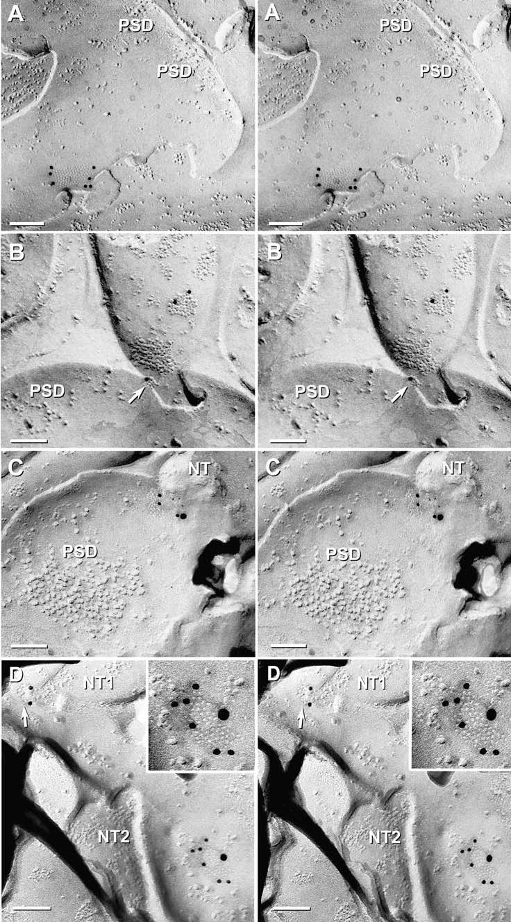
High-magnification stereoscopic images of Cx36 single-labeled and Cx36 and Cx45 double-labeled gap junctions at “mixed” synapses in adult mouse olfactory bulb. (A) E-face image of neuronal gap junction labeled for Cx36 (12 nm gold beads). PSD = postsynaptic density. (B) Two Cx36-labeled gap junctions on a presynaptic neurite. Note the narrowing of the extracellular space at the point of contact of the lower gap junction (arrow). PSD = postsynaptic density. (C) Gap junction double-labeled for Cx36 (12 nm gold beads) and Cx45 (6 nm and 18 nm gold beads). NT = nerve terminal. PSD = postsynaptic density. (D) Two gap junctions linking two neurites (NT1 and NT2) to a postsynaptic dendrite. Upper gap junction (arrow) is single-labeled for Cx36, whereas the lower gap junction (shown at higher magnification in the inset) is double labeled for Cx36 (six 12 nm gold beads) and Cx45 (one 18 nm gold bead).