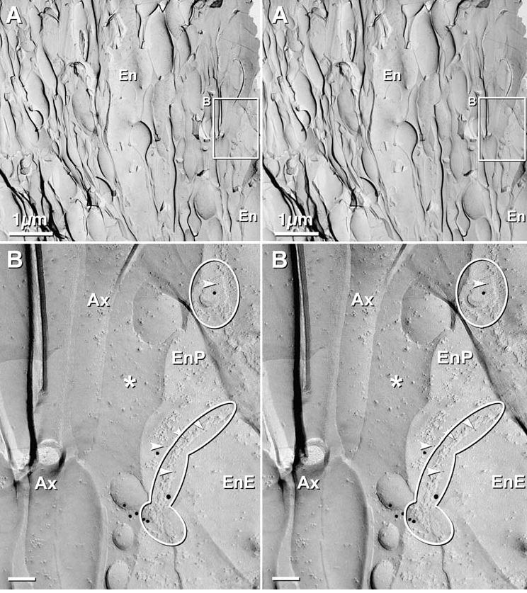
Fig. 8.

Stereoscopic images of axon bundle and surrounding ensheathing cells in OE of 4 day postnatal rat after double-labeling for Cx36 and Cx43, using 5 sizes of gold (Cx36 = 10 nm and 12 nm; Cx43 = 5 nm, 18 nm, and 20 nm). (A) Low magnification image of an axon bundle, with an intrafascicular ensheathing cell (En, top center) and an external ensheathing cell (En at lower right; and inscribed box, shown at higher magnification below). (B) Higher magnification stereoscopic image of the inscribed area “B”, above. Two ensheathing cell gap junctions are delineated by ovals. Each gap junction is labeled for Cx43, with 5 nm, 18 nm and 20–22 nm gold beads. No 10 nm or 12 nm gold beads (for Cx36) are present on gap junctions in ensheathing cells or in axons. The small clump of 18nm gold beads (bottom center) was counted as a single “hit” (Rash and Yasumura, 1999). * = extracellular space; Arrowheads = 5 nm gold beads; Ax = axon; EnE = ensheathing cell E-face; EnP = ensheathing cell P-face.
