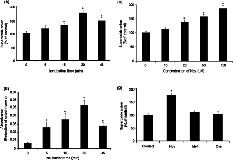
Figure 1. Superoxide anion levels in monocytes.
Cells were incubated with or without Hcy (100 μM) for various time periods. At the end of the incubation, the intracellular superoxide anion was measured by NBT reduction assay (A) and the extracellular superoxide anion was measured using cytochrome c reduction assay (B). (C) Cells were incubated with Hcy (10–100 μM) or without Hcy for 30 min. (D) Cells were incubated for 30 min in the absence (Control) or presence of Hcy (100 μM), cysteine (Cys; 100 μM) or methionine (Met; 100 μM). In (C and D), the levels of superoxide anions in these cells were determined by NBT reduction assay. The level of superoxide anions in control cells was expressed as 100%. Results were expressed as means±S.E.M. for five separate experiments each performed in duplicate. *Significantly different from the control value obtained from cells incubated in the absence of Hcy (P<0.05).

