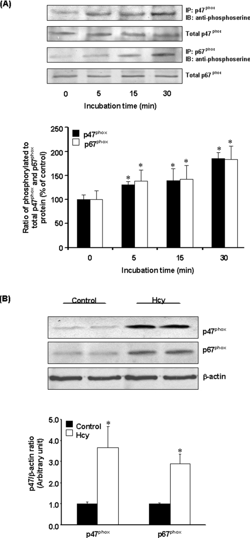
Figure 5. Phosphorylation of p47phox and p67phox subunits.
(A) Cells were incubated with Hcy (100 μM) or without Hcy for various time periods. p47phox and p67phox proteins were immunoprecipitated (IP) using goat anti-p47phox or p67phox antibodies followed by Western-blot analysis (IB) using anti-phosphoserine antibodies to detect serine-phosphorylated proteins. The immunoblots were analysed by densitometry. Results were normalized to total p47phox and p67phox proteins in the immunoprecipitates and are means±S.E.M. for five separate experiments. The relative densitometric units for the ratio of phosphorylated to total p47phox or p67phox proteins in the control cells (incubated in the absence of Hcy) were expressed as 100%. (B) Cells were incubated for 30 min in the absence (Control) or presence of Hcy (100 μM). The membrane fraction was prepared for detection of p47phox or p67phox proteins by Western-blot analysis. β-Actin was used to confirm equal amount of protein loading for each sample. Representative blots were obtained from five separate experiments. *Significantly different from the control value obtained from cells incubated in the absence of Hcy (P<0.05).

