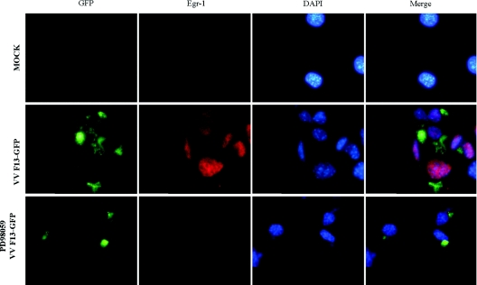
Figure 4. VV stimulates EGR-1 nuclear translocation.
Immunofluorescence microscopy of EGR-1 cellular localization in virus-infected cells. Cells growing on coverslips were mock-infected (top row) or infected with VVF13-GFP (green) at an MOI of 10.0 for 12 h and then stained with anti-EGR-1 antibody (red), either in the absence (middle row) or in the presence of PD98059 (50 μM) (bottom row). Nuclei were stained with DAPI (4′,6-diamidino-2-phenylindole). Merged pictures show the DAPI-stained image superimposed on the EGR-1-stained image. Results were confirmed by three independent assays with similar results.

